โรคของซีลิแอ็ก
| โรคของซีลิแอ็ก (Coeliac disease) | |
|---|---|
| ชื่ออื่น | Celiac sprue, nontropical sprue, endemic sprue, gluten enteropathy |
 | |
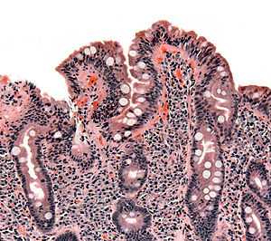
| เนื้อที่ตัดจากลำไส้เล็กออกตรวจ แสดงลักษณะของ coeliac disease คือมีส่วนยื่นที่ทื่อลง (blunting of villi) ส่วนม้วนเข้าที่ใหญ่เกิน (crypt hypertrophy) และลิมโฟไซต์เข้าไปในส่วนม้วนเข้า (lymphocyte infiltration of crypt) | |
| การออกเสียง |
|
| สาขาวิชา | วิทยาทางเดินอาหาร อายุรศาสตร์ |
| อาการ | ไม่มีหรือไม่เจำเพาะ, ท้องยื่น/ท้องพอง, ท้องร่วง, ท้องผูก, ดูดซึมอาหารไม่ดี, น้ำหนักลด, dermatitis herpetiformis[A][5][6] |
| ภาวะแทรกซ้อน | ภาวะเลือดจางเหตุขาดธาตุเหล็ก ภาวะกระดูกพรุน เป็นหมัน มะเร็งต่าง ๆ ปัญหาทางประสาท และโรคภูมิต้านตนเองอื่น ๆ[7][8][9][10][11] |
| การตั้งต้น | ไม่จำกัดอายุ[5][12] |
| ระยะดำเนินโรค | เป็นตลอดชีวิต[10] |
| สาเหตุ | ปฏิกิริยาต่อกลูเตน[13] |
| วิธีวินิจฉัย | สอบประวัติครอบครัว ตรวจสารภูมิต้านทานในเลือด ตัดเนื้อลำไส้ออกตรวจ ตรวจพันธุกรรม ดูการตอบสนองเมื่องดกลูเตน[14][15] |
| โรคอื่นที่คล้ายกัน | ลำไส้อักเสบ พยาธิลำไส้ กลุ่มอาการลำไส้ไวเกินต่อการกระตุ้น ซิสติกไฟโบรซิส[16] |
| การรักษา | อาหารปลอดกลูเตน[17] |
| ความชุก | ~1 คนใน 135 คน[18] |
โรคของซีลิแอ็ก[19] (อังกฤษ: coeliac disease หรือ celiac disease) เป็นโรคภูมิต้านตนเองระยะยาวซึ่งสร้างปัญหาโดยหลักต่อลำไส้เล็ก[14] อาการตามแบบรวมทั้งปัญหากระเพาะลำไส้ เช่น ท้องร่วงเรื้อรัง ท้องพอง/ท้องยื่น (abdominal distention) ดูดซึมอาหารไม่ดี (malabsorption) ไม่อยากอาหาร และไม่โต (ในเด็ก)[5] ปกติจะเกิดเมื่ออายุระหว่าง 6 เดือนถึง 2 ขวบ[5] แต่อาการอื่น ๆ จะสามัญกว่าโดยเฉพาะในผู้มีอายุมากกว่า 2 ปี[12][20][21][22] อาการกระเพาะลำไส้อาจจะเบาหรือไม่มี อาจมีอาการที่ส่วนอื่น ๆ ของร่างกาย หรือไม่ปรากฏอาการอะไร ๆ เลย[5] แม้โรคอาจเริ่มตั้งแต่วัยเด็ก[10][12] แต่ก็เกิดเมื่ออายุเท่าไรก็ได้[5][12] โรคมักสัมพันธ์กับโรคภูมิต้านตนเองอื่น ๆ เช่น โรคเบาหวานประเภทที่ 1 และต่อมไทรอยด์อักเสบเป็นต้น[10]
โรคเกิดจากปฏิกิริยาของร่างกายต่อกลูเตน ซึ่งเป็นโปรตีนต่าง ๆ ที่พบในข้าวสาลีและข้าวประเภทอื่น ๆ รวมทั้งข้าวบาร์เลย์และข้าวไรย์[13][23][24] ปกติคนไข้สามารถรับข้าวโอ๊ตที่ไม่มากเกินโดยไม่เจือปนกับข้าวที่มีกลูเตนอื่น ๆ ได้[23][25] แต่ก็อาจมีปัญหากับข้าวโอ๊ตบางสายพันธุ์[23][26] เป็นโรคที่เกิดกับผู้มีปัญหาทางพันธุกรรม[14] คือ เมื่อได้รับกลูเตน การตอบสนองผิดปกติของระบบภูมิคุ้มกันจะผลิตสารภูมิต้านทานต้านตนเอง (autoantibody) ซึ่งอาจสร้างปัญหาแก่อวัยวะต่าง ๆ[8][27] ในลำไส้เล็ก นี่ทำให้อักเสบ และอาจทำส่วนยื่นที่บุลำไส้ (intestinal villus) ให้สั้น/ทื่อลง เป็นอาการที่เรียกว่า villous atrophy[14][15] ซึ่งทำให้ดูดซึมอาหารได้น้อยลง และบ่อยครั้งทำให้โลหิตจาง[14][24]
การวินิจฉัยจะอาศัยการตรวจสารภูมิต้านทานในเลือดบวกกับการตัดเนื้อลำไส้ออกตรวจ โดยการทดสอบทางพันธุกรรม (genetic testing) โดยเฉพาะอาจช่วย[14] แต่บางครั้งก็วินิจฉัยได้ยาก[28] เพราะอาจตรวจไม่พบสารภูมิต้านทานต้านตนเอง (autoantibody)[29][30] เพราะลำไส้เล็กอาจเปลี่ยนไปเพียงเล็กน้อยและส่วนยื่นจะดูปกติ[21][31] แม้ผู้ที่มีอาการหนักก็อาจต้องไปหาหมอเป็นเวลาหลายปีกว่าจะวินิจฉัยได้ถูกต้อง[32] ปัจจุบันมีผู้ไม่มีอาการอะไร ๆ ที่วินิจฉัยว่ามีโรคนี้ โดยอาศัยการตรวจคัดโรค[33] แต่หลักฐานซึ่งแสดงประโยชน์ของการตรวจคัดโรคก็ยังไม่ชัดเจน[34] แม้โรคจะมีเหตุจากความไม่ทนต่อโปรตีนข้าวสาลี แต่นี่ก็ไม่ใช่ภาวะภูมิแพ้ข้าวสาลี (wheat allergy)[14]
วิธีการรักษาในปัจจุบันอย่างเดียวก็คือการให้ทานอาหารปลอดกลูเตนตลอดชีวิต ซึ่งอาจทำให้เยื่อลำไส้ฟื้นตัว ทำให้อาการดีขึ้น และลดความเสี่ยงภาวะแทรกซ้อนอื่น ๆ ซึ่งได้ผลต่อคนไข้โดยมาก[17] ถ้าไม่รักษา นี่อาจทำให้เกิดมะเร็ง เช่น มะเร็งต่อมน้ำเหลืองของลำไส้ (intestinal lymphoma) และเพิ่มความเสี่ยงการตายก่อนวัยโดยเล็กน้อย[7] อัตราการเกิดโรคไม่เหมือนกันในเขตต่าง ๆ ทั่วโลก เริ่มจากความชุกโรคที่ 1 คนต่อ 300 คนจนถึง 1 คนต่อ 40 คน โดยเฉลี่ยที่ระหว่าง 1 คนต่อ 100 คน กับ 1 คนใน 170 คน[18] ในประเทศพัฒนาแล้ว ประเมินว่า คนไข้ 80% ยังไม่ได้วินิจฉัย เพราะมีปัญหากระเพาะลำไส้น้อยมากหรือไม่มีเลย และเพราะไม่รู้เรื่องโรค[9][35] เป็นโรคที่สามัญในหญิงมากกว่าชาย[36] คำภาษาอังกฤษว่า coeliac มาจากคำภาษากรีกว่า κοιλιακός (koiliakós, "abdominal" แปลว่า เกี่ยวกับท้อง) โดยเริ่มใช้ตั้งแต่คริสต์ศตวรรษที่ 19 อาศัยงานแปลจากภาษากรีกโบราณเกี่ยวกับโรคของแพทย์ชาวกรีกคือ Aretaeus of Cappadocia[37][38]
เชิงอรรถ
[แก้]อ้างอิง
[แก้]- ↑ Freedberg; และคณะ (2003). Fitzpatrick's Dermatology in General Medicine (6th ed.). McGraw-Hill. ISBN 0-07-138076-0.
- ↑ Rapini, Ronald P.; Bolognia, Jean L.; Jorizzo, Joseph L. (2007). Dermatology. St. Louis: Mosby. ISBN 1-4160-2999-0.
- ↑ Singal A, Bhattacharya SN, Baruah MC (2002). "Dermatitis herpetiformis and rheumatoid arthritis". Indian J Dermatol Venereol Leprol. 68 (4): 229–30. PMID 17656946.
- ↑ "Dermatitis Herpetiformis". American Osteopathic College of Dermatology. เก็บจากแหล่งเดิมเมื่อ 2018-10-29.
- ↑ 5.0 5.1 5.2 5.3 5.4 5.5 Fasano, A (April 2005). "Clinical presentation of celiac disease in the pediatric population". Gastroenterology (Review). 128 (4 Suppl 1): S68-73. doi:10.1053/j.gastro.2005.02.015. PMID 15825129.
- ↑ "Symptoms & Causes of Celiac Disease | NIDDK". National Institute of Diabetes and Digestive and Kidney Diseases. June 2016. เก็บจากแหล่งเดิมเมื่อ 2017-04-24. สืบค้นเมื่อ 2017-04-24.
- ↑ 7.0 7.1 Lebwohl B, Ludvigsson JF, Green PH (October 2015). "Celiac disease and non-celiac gluten sensitivity". BMJ (Review). 351: h4347. doi:10.1136/bmj.h4347. PMC 4596973. PMID 26438584.
Celiac disease occurs in about 1% of the population worldwide, although most people with the condition are undiagnosed. It can cause a wide variety of symptoms, both intestinal and extra-intestinal because it is a systemic autoimmune disease that is triggered by dietary gluten. Patients with coeliac disease are at increased risk of cancer, including a twofold to fourfold increased risk of non-Hodgkin’s lymphoma and a more than 30-fold increased risk of small intestinal adenocarcinoma, and they have a 1.4-fold increased risk of death.
- ↑ 8.0 8.1 Lundin KE, Wijmenga C (September 2015). "Coeliac disease and autoimmune disease-genetic overlap and screening". Nature Reviews. Gastroenterology & Hepatology (Review). 12 (9): 507–15. doi:10.1038/nrgastro.2015.136. PMID 26303674.
The abnormal immunological response elicited by gluten-derived proteins can lead to the production of several different autoantibodies, which affect different systems.
- ↑ 9.0 9.1 "Celiac disease". World Gastroenterology Organisation Global Guidelines. July 2016. เก็บจากแหล่งเดิมเมื่อ 2017-03-17. สืบค้นเมื่อ 2017-04-23.
- ↑ 10.0 10.1 10.2 10.3
Ciccocioppo R, Kruzliak P, Cangemi GC, Pohanka M, Betti E, Lauret E, Rodrigo L (2015-10-22). "The Spectrum of Differences between Childhood and Adulthood Celiac Disease". Nutrients (Review). 7 (10): 8733–51. doi:10.3390/nu7105426. PMC 4632446. PMID 26506381.
Several additional studies in extensive series of coeliac patients have clearly shown that TG2A sensitivity varies depending on the severity of duodenal damage, and reaches almost 100% in the presence of complete villous atrophy (more common in children under three years), 70% for subtotal atrophy, and up to 30% when only an increase in IELs is present. (IELs: intraepithelial lymphocytes)
- ↑
Lionetti E, Francavilla R, Pavone P, Pavone L, Francavilla T, Pulvirenti A, Giugno R, Ruggieri M (August 2010). "The neurology of coeliac disease in childhood: what is the evidence? A systematic review and meta-analysis". Developmental Medicine and Child Neurology. 52 (8): 700–7. doi:10.1111/j.1469-8749.2010.03647.x. PMID 20345955.

- ↑ 12.0 12.1 12.2 12.3
Husby S, Koletzko S, Korponay-Szabó IR, Mearin ML, Phillips A, Shamir R, Troncone R, Giersiepen K, Branski D, Catassi C, Lelgeman M, Mäki M, Ribes-Koninckx C, Ventura A, Zimmer KP, ESPGHAN Working Group on Coeliac Disease Diagnosis, ESPGHAN Gastroenterology Committee, European Society for Pediatric Gastroenterology, Hepatology, Nutrition (January 2012). "European Society for Pediatric Gastroenterology, Hepatology, and Nutrition guidelines for the diagnosis of coeliac disease" (PDF). J Pediatr Gastroenterol Nutr (Practice Guideline). 54 (1): 136–60. doi:10.1097/MPG.0b013e31821a23d0. PMID 22197856. เก็บ (PDF)จากแหล่งเดิมเมื่อ 2016-04-03.
Since 1990, the understanding of the pathological processes of CD has increased enormously, leading to a change in the clinical paradigm of CD from a chronic, gluten-dependent enteropathy of childhood to a systemic disease with chronic immune features affecting different organ systems. (...) atypical symptoms may be considerably more common than classic symptoms
- ↑ 13.0 13.1 Tovoli F, Masi C, Guidetti E, Negrini G, Paterini P, Bolondi L (March 2015). "Clinical and diagnostic aspects of gluten related disorders". World Journal of Clinical Cases (Review). 3 (3): 275–84. doi:10.12998/wjcc.v3.i3.275. PMC 4360499. PMID 25789300.
- ↑ 14.0 14.1 14.2 14.3 14.4 14.5 14.6 "Celiac Disease". NIDDKD. June 2015. เก็บจากแหล่งเดิมเมื่อ 2016-03-13. สืบค้นเมื่อ 2016-03-17.
- ↑ 15.0 15.1 Vivas S, Vaquero L, Rodríguez-Martín L, Caminero A (November 2015). "Age-related differences in celiac disease: Specific characteristics of adult presentation". World Journal of Gastrointestinal Pharmacology and Therapeutics (Review). 6 (4): 207–12. doi:10.4292/wjgpt.v6.i4.207. PMC 4635160. PMID 26558154.
In addition, the presence of intraepithelial lymphocytosis and/or villous atrophy and crypt hyperplasia of small-bowel mucosa, and clinical remission after withdrawal of gluten from the diet, are also used for diagnosis antitransglutaminase antibody (tTGA) titers and the degree of histological lesions inversely correlate with age. Thus, as the age of diagnosis increases antibody titers decrease and histological damage is less marked. It is common to find adults without villous atrophy showing only an inflammatory pattern in duodenal mucosa biopsies: Lymphocytic enteritis (Marsh I) or added crypt hyperplasia (Marsh II)
- ↑ Ferri, Fred F. (2010). Ferri's differential diagnosis : a practical guide to the differential diagnosis of symptoms, signs, and clinical disorders (2nd ed.). Philadelphia, PA: Elsevier/Mosby. p. Chapter C. ISBN 0323076998.
- ↑ 17.0 17.1 See JA, Kaukinen K, Makharia GK, Gibson PR, Murray JA (October 2015). "Practical insights into gluten-free diets". Nature Reviews. Gastroenterology & Hepatology (Review). 12 (10): 580–91. doi:10.1038/nrgastro.2015.156. PMID 26392070.
A lack of symptoms and/or negative serological markers are not reliable indicators of mucosal response to the diet. Furthermore, up to 30% of patients continue to have gastrointestinal symptoms despite a strict GFD.122,124 If adherence is questioned, a structured interview by a qualified dietitian can help to identify both intentional and inadvertent sources of gluten.
- ↑ 18.0 18.1 Fasano A, Catassi C (December 2012). "Clinical practice. Celiac disease". The New England Journal of Medicine (Review). 367 (25): 2419–26. doi:10.1056/NEJMcp1113994. PMID 23252527.
- ↑ บัญชีจำแนกโรคระหว่างประเทศ ฉบับประเทศไทย (อังกฤษ-ไทย) ฉบับปี 2009. สำนักนโยบายและยุทธศาสตร์ สำนักงานปลัดกระทรวงสาธารณสุข, 2552.
- ↑
Newnham, Evan D (2017). "Coeliac disease in the 21st century: Paradigm shifts in the modern age". Journal of Gastroenterology and Hepatology. 32: 82–85. doi:10.1111/jgh.13704. PMID 28244672.
Presentation of CD with malabsorptive symptoms or malnutrition is now the exception rather than the rule.

- ↑ 21.0 21.1 M, Rostami Nejad; Hogg-Kollars, S; Ishaq, S; Rostami, K (2011). "Subclinical celiac disease and gluten sensitivity". Gastroenterol Hepatol Bed Bench (Review). 4 (3): 102–8. PMC 4017418. PMID 24834166.
- ↑ Tonutti E, Bizzaro N (2014). "Diagnosis and classification of celiac disease and gluten sensitivity". Autoimmun Rev (Review). 13 (4–5): 472–6. doi:10.1016/j.autrev.2014.01.043. PMID 24440147.
- ↑ 23.0 23.1 23.2 Penagini F, Dilillo D, Meneghin F, Mameli C, Fabiano V, Zuccotti GV (November 2013). "Gluten-free diet in children: an approach to a nutritionally adequate and balanced diet". Nutrients (Review). 5 (11): 4553–65. doi:10.3390/nu5114553. PMC 3847748. PMID 24253052.
- ↑ 24.0 24.1 Di Sabatino A, Corazza GR (April 2009). "Coeliac disease". Lancet. 373 (9673): 1480–93. doi:10.1016/S0140-6736(09)60254-3. PMID 19394538.
- ↑ Pinto-Sánchez MI, Causada-Calo N, Bercik P, Ford AC, Murray JA, Armstrong D, Semrad C, Kupfer SS, Alaedini A, Moayyedi P, Leffler DA, Verdú EF, Green P (August 2017). "Safety of Adding Oats to a Gluten-Free Diet for Patients With Celiac Disease: Systematic Review and Meta-analysis of Clinical and Observational Studies". Gastroenterology. 153 (2): 395-409.e3. doi:10.1053/j.gastro.2017.04.009. PMID 28431885.
- ↑ Comino I, Moreno M, Sousa C (November 2015). "Role of oats in celiac disease". World Journal of Gastroenterology. 21 (41): 11825–31. doi:10.3748/wjg.v21.i41.11825. PMC 4631980. PMID 26557006.
It is necessary to consider that oats include many varieties, containing various amino acid sequences and showing different immunoreactivities associated with toxic prolamins. As a result, several studies have shown that the immunogenicity of oats varies depending on the cultivar consumed. Thus, it is essential to thoroughly study the variety of oats used in a food ingredient before including it in a gluten-free diet.
- ↑ National Institute for Health and Clinical Excellence. Clinical guideline 86: Recognition and assessment of coeliac disease. London, 2015.
- ↑ Matthias T, Pfeiffer S, Selmi C, M Eric Gershwin (April 2010). "Diagnostic challenges in celiac disease and the role of the tissue transglutaminase-neo-epitope". Clin Rev Allergy Immunol (Review). 38 (2–3): 298–301. doi:10.1007/s12016-009-8160-z. PMID 19629760.
- ↑ Lewis NR, Scott BB (July 2006). "Systematic review: the use of serology to exclude or diagnose coeliac disease (a comparison of the endomysial and tissue transglutaminase antibody tests)". Alimentary Pharmacology & Therapeutics. 24 (1): 47–54. doi:10.1111/j.1365-2036.2006.02967.x. PMID 16803602.
- ↑ Rostom A, Murray JA, Kagnoff MF (December 2006). "American Gastroenterological Association (AGA) Institute technical review on the diagnosis and management of celiac disease". Gastroenterology (Review). 131 (6): 1981–2002. doi:10.1053/j.gastro.2006.10.004. PMID 17087937.
- ↑ Molina-Infante J, Santolaria S, Sanders DS, Fernández-Bañares F (May 2015). "Systematic review: noncoeliac gluten sensitivity". Alimentary Pharmacology & Therapeutics (Review). 41 (9): 807–20. doi:10.1111/apt.13155. PMID 25753138.
Furthermore, seronegativity is more common in coeliac disease patients without villous atrophy (Marsh 1-2 lesions), but these ‘minor’ forms of coeliac disease may have similar clinical manifestations to those with villous atrophy and may show similar clinical-histological remission with reversal of haematological or biochemical disturbances on a gluten-free diet (GFD).
- ↑ Ludvigsson JF, Card T, Ciclitira PJ, Swift GL, Nasr I, Sanders DS, Ciacci C (April 2015). "Support for patients with celiac disease: A literature review". United European Gastroenterology Journal (Review). 3 (2): 146–59. doi:10.1177/2050640614562599. PMC 4406900. PMID 25922674.
- ↑ van Heel DA, West J (July 2006). "Recent advances in coeliac disease". Gut (Review). 55 (7): 1037–46. doi:10.1136/gut.2005.075119. PMC 1856316. PMID 16766754.
- ↑ Bibbins-Domingo K, Grossman DC, Curry SJ, Barry MJ, Davidson KW, Doubeni CA, Ebell M, Epling JW, Herzstein J, Kemper AR, Krist AH, Kurth AE, Landefeld CS, Mangione CM, Phipps MG, Silverstein M, Simon MA, Tseng CW (March 2017). "Screening for Celiac Disease: US Preventive Services Task Force Recommendation Statement". JAMA. 317 (12): 1252–1257. doi:10.1001/jama.2017.1462. PMID 28350936.
- ↑ Lionetti E, Gatti S, Pulvirenti A, Catassi C (June 2015). "Celiac disease from a global perspective". Best Practice & Research. Clinical Gastroenterology (Review). 29 (3): 365–79. doi:10.1016/j.bpg.2015.05.004. PMID 26060103.
- ↑ Hischenhuber C, Crevel R, Jarry B, Mäki M, Moneret-Vautrin DA, Romano A, Troncone R, Ward R (March 2006). "Review article: safe amounts of gluten for patients with wheat allergy or coeliac disease". Alimentary Pharmacology & Therapeutics. 23 (5): 559–75. doi:10.1111/j.1365-2036.2006.02768.x. PMID 16480395.
- ↑ Adams F (1856). "On The Cœliac Affection". The extant works of Aretaeus, The Cappadocian. London: Sydenham Society. pp. 350–1. สืบค้นเมื่อ 2009-12-12.
- ↑ Losowsky, MS (2008). "A history of coeliac disease". Digestive Diseases. 26 (2): 112–20. doi:10.1159/000116768. PMID 18431060.
แหล่งข้อมูลอื่น
[แก้]- โรคของซีลิแอ็ก ที่เว็บไซต์ Curlie
| การจำแนกโรค | |
|---|---|
| ทรัพยากรภายนอก |
